
Digital Imaging
We have given numerous talks at major medical conferences including the Keynote at the Visions conference. Our pathologists also participated in the 510K FDA clearance of breast markers.
Affiliated Pathologists Medical Group recently joined the Professional Network of Xifinl. The organization is a comprehensive online information exchange and digital consultation forum. It gives pathologists around the world access to our expertise. You can read more about it HERE.
Uses
We have expanded the use of whole slide imaging into a number of niche applications: tumor board presentations, quality assurance, intra and extra departmental consultations, virtual immunohistochemistry, and virtual slide box, archiving of rare or medical legal cases, and digital slide consultations with clients. Scanned slides can also be shared for consultations with subspecialty experts within our group.
A few academic medical centers are establishing WSI outsourcing programs. Our association with PathCentral gives APMG this capability as well. You can read more about our insourcing program on this page and our client consultation program on this page. We've also listed digital pathology resources on this page.
Three other high-impact uses are highlighted below.
Tumor Conference Presentations
There is absolutely no better way for a pathologist to present tumor conference cases than whole slide images. The process is engaging, dynamic, and interactive. Clinicians get a real sense of what the case is all about and how the diagnosis was rendered. Time and time again we get compliments for our tumor conference presentations. Scripps University recently conducted a satisfaction survey and our experience matches exactly.
Virtual Immunohistochemistry and Image Analysis
We are partners with reference laboratories in Southern California that use whole slide imaging and are available for clinical trials, reference work, virtual IHC studies, and image analyses.
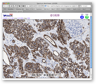
One of the primary drivers for setting up whole slide imaging was virtual immunohistochemistry. We have an outstanding immunohistochemistry department at Pathology, Inc. that provides stains to universities, large medical centers, and pathologists at community hospitals. Glass slides are returned via courier or UPS but the referring pathologists can view the results the same day they are performed using digitally scanned slides. This improves turn-around-time and allows ordering a second set of stains immediately, if needed.
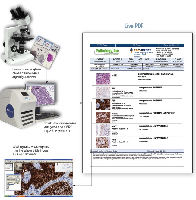
Image analysis is the application of computer-assisted algorithms to the whole slide image. The most common use is for breast markers—ER, PR, HER2, and Ki67. APMG pathologists perform the analysis using the ASCO-CAP guidelines.
The evaluation of the whole slide image can be set up as "tech only" for those pathologists who wish to perform and bill for their own evaluations.
The final report is a PDF, as shown below. What is unique is that each photographic image is a live link to the respective whole slide image. This allows the pathologist to directly view the digital slide by simply clicking on the photo in the PDF (after entering a name and password). We call this capability Live PDF and it is one more innovative differentiator for APMG.
More information can be found on these pages: Remote Consultations, Realtime Consultations, and Resources.